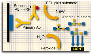

The antigen (in our case Cancer Antigen 1 [CA1] {blue rectangle}) is bound to the nitrocellulose paper (thick black line). First we expose the nitrocellulose paper to a solution containing the specific antibody against CA1 (the primary antibody). We then expose the nitrocellulose paper to the secondary antibody which is conjugated to the enzyme HRP - a peroxidase {red circle}. The secondary antibody binds to the primary antibody which is bound to the antigen (CA1). We now immerse the nitrocellulose paper in a buffer containing the substrate of the peroxidase and then transfer it on top of a film for a short period (seconds to minutes). The peroxidase (which is found only where the antigen [CA1] is located) catalyses a light emitting reaction which is detected on the film.