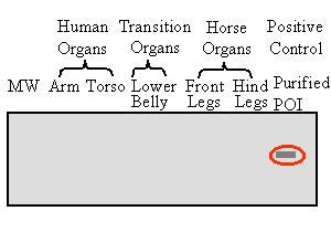
1. Wrong
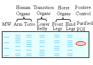
- we can easily recognize the purified POI (our positive control) in the
last sample to the right, where it is the only protein in the sample (circled
in red). Indeed, all other samples contain bands of
simillar size. However, these bands can be positively identified as POI,
only if they are recognised by the POI specific antibody, used in the Western
blot (lower panel). Since these bands are not
recognized by the POI specific antigen, they are unrelated to POI though
they have a simillar size. Indeed, whole cell lysates contain numerous
proteins of various quantities, some of which may be of the same size.
Commassie staining is less sensitive than immunoblotting. It detects higher amounts of ALL proteins. |
|
Possible but less likely - the purified POI (our positive control) is visualized (circled in red). Thus, we can safely conclude that the primary antibody, the secondary antibody and the reaction catalysed by the reporter enzyme - all work well. The only thing that could go wrong is the transfer of proteins from the gel to the nitrocellulose membrane. Lack of buffer or presence of air bubbles in the left part of the apparatus could theoretically explain the detection of POI samples in part of the gel. However, this explanation is less likely. |
3. Right
- we should use more subtle dissection methods to increase the relative
amounts of POI in our samples.
Alternatively, we can take
the whole cell lysates, obtained as previously described, and perform immunoprecipitation
using anti-POI antibodies to increase the relative amounts of the POI protein
in our sample before we perfom immunoblotting.
.