 How many copies of the studied gene do you expect to
find in each sample?
How many copies of the studied gene do you expect to
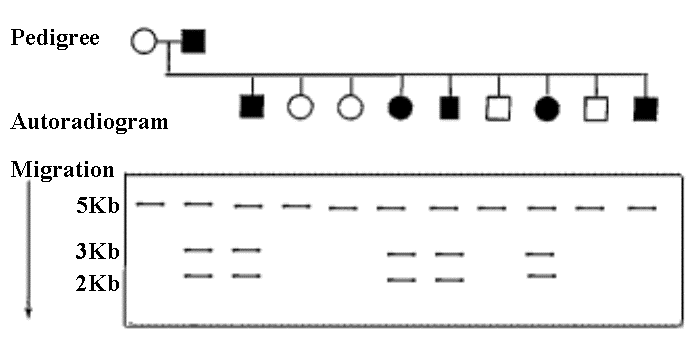
find in each sample?DNA studies are performed on a large family that shows a certain autosomal dominant disease of late onset (approximately 40 years of age). A DNA sample from each family member is digested with the restriction enzyme TaqI and subjected to gel electrophoresis. A Southern blot is then performed, with the use of a long radioactive probe consisting of part of the studied gene .
 How many copies of the studied gene do you expect to
find in each sample?
How many copies of the studied gene do you expect to
find in each sample?The family pedigree and the Southern blot autoradiogram are shown below. Affected members are shown in black.

 What
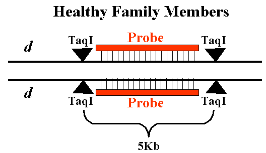
is the DNA restriction profile of the healthy family members (shown in
white)?
 |
How can you explain the case of the last son who is affected by the disease but has only the 5Kb DNA fragment? |

This is an autosomal disease,
so both males and females are expected to have two copies of the studied
gene. However, the healthy family members have the dd allele
combination, whereas the affected family members have the Dd allele
combination.
|
|
|
All healthy family members
(dd) have only the 5Kb band. The probe hybridizes to a sequence
~ 5Kb between the two TaqI recognition sites. (The probe can
also slightly protrude beyond one of the TaqI cleavage sites.)
|
 |
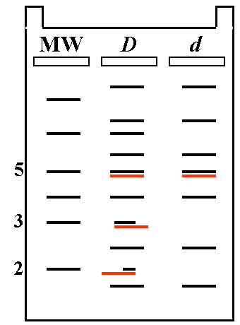
| Allaffected family members have the Dd allele combination. The D allele bears an additional TaqI site in most cases, resulting in the formation of two smaller fragments of 2Kb and 3Kb, instead of the 5Kb fragment characteristic of the d allele. |  |
||
| Since the probe
used is long enough, it can still hybridize with the two shorter TaqI
fragments of the D allele under appropriate conditions.
|
 |
|
|
|
|
|
|
.